Dr LEE Ming-Yueh 李明月医生

 Glaucoma    Types of Glaucoma    Glaucoma Tests    Glaucoma Treatment Options    Glaucoma Cases  

Glaucoma Tests


A. Tonometry

Eye pressure can be measured by few methods. It can be measured either by contact or non-contact tonometer. The most accurate and the gold standard of all is Goldman Tonometer. It is commonly used by eye doctors worldwide.

Tonometry

Fig: Measuring IOP with Goldman Tonometer. 
*image obtained from google images


B. Slit Lamp Examination

Slit lamp is a high tech instrument with magnifiying lens used by eye doctors to do detail examination of the eye. It can be used with additional hand-held magnifying lens to get a clear view of the optic nerve, macula and retina.

Tonometry

Fig: Dr Lee examined a patient’s eye with Slit Lamp. 
*picture taken during public screening on World Glaucoma Day 2014


C. Fundus Photography

Optic disc image can be captured with fundus camera for documentation and for monitoring of progress of the disease.


images/Glaucoma Optic Disc
Fig: Normal Optic Disc (Optic Nerve Head) with Cup-Disc ratio of 0.3
Fig: Glaucoma Optic Disc with advanced nerve damage Cup-Disc ratio of 0.9
The pale area around the Optic Disc is a 
result of death of optic nerve cells


D. Gonioscopy

Gonioscopy is a procedure where a Gonioslens is used to visualize the angle structure of the eye.

Gonioscopy is necessary for grading of the angle and and to look for any causes that obstruct the fluid/aquoeus outflow. This is an important procedure to help glaucoma specialists to come to a diagnosis/type of glaucoma that you have.


Angle structure of the eye
Khaw Goniolens
Fig: Drawing of Angle structure of the eye.
*image obtained from google images
Fig: Khaw Goniolens
*image obtained from google images
   
Gonioscopy view of Eye with Open Angle
Gonioscopy view of Eye with Narrow Angle
Fig: Gonioscopy view of Eye with Open Angle
*image obtained from google images
Fig: Gonioscopy view of Eye with Narrow Angle
*image obtained from google images


E. Visual Field Test

Humprey Visual Field test is the gold standard of visual field test for glaucoma. It helps to map-out the visual field defect of a glaucoma patient.

A patient could not tell how much visual vield he has lost because he himself does not aware of it not until the defect is being maped-out by this computer-assisted machine.


Fig: Progressive visual field loss with corresponding constricting view by a glaucoma patient 
*image obtained from google images


Video from http://eyewiki.aao.org


F. Cornea Thickness

The normal cornea thickness is around 520 -530 um. Studies showed patient with thin cornea have higher risk of glaucoma. The information of cornea thickness is important for glaucoma specialists  to plan your treatment and to make a decision on your target eye pressure (IOP) to prevent further optic nerve damage.


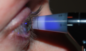
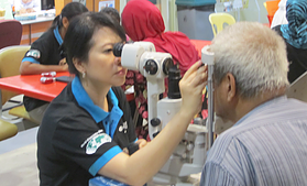
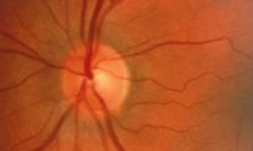
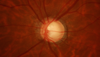
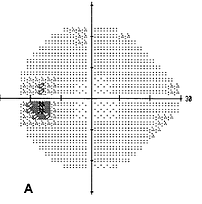
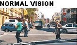
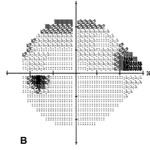
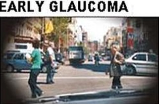
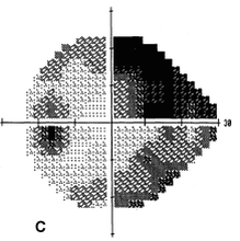
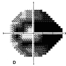
G. Retinal Nerve Fibre Layer Assessment

Retinal nerve fibre layer could be analysed by using the following diagnostic instruments. These tests are useful as supplimentary for early diagnosis and monitoring progression of glaucoma.

1. Optical Coherence Tomography

2. Heidelberg Retinal Tomography (HRT)

3. Scanning Laser Polarimeter (GDx)